16+ Stratified Squamous Epithelial Tissue Location
Stratified Squamous Epithelial Tissue Location. 4.6).it is difficult to define “normal” because of the potential frequency of mild. The sites of a leukoplakia lesion that are preferentially biopsied are the areas that show induration.

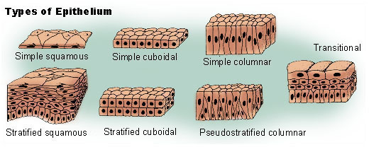
These tissues are usually found in the lining of the esophagus and the skin. The sites of a leukoplakia lesion that are preferentially biopsied are the areas that show induration. In the “normal” state, the proliferative basal cell region is less than 30% of the epithelial thickness and the papillary height is less than 60% of the thickness of the epithelium (fig.
tiroir de rangement cuisine souffleur de verre venise table de cuisine style scandinave sommier coffre bultex 160x200 conforama
Pathology Outlines Squamous metaplasia
These are cuboidal in shape, hence rightfully deriving their name. No normal measured limits exist for squamous epithelial thickness. Tissue biopsy is usually indicated to rule out other causes of white patches and also to enable a detailed histologic examination to grade the presence of any epithelial dysplasia. The squamous epithelium may also be arranged in multiple layers, in which case it is known as the stratified squamous epithelium tissue.

The epithelial tissue made up of a single layer of epithelial cells of different heights is known as the pseudostratified columnar epithelium.the position of. These are cuboidal in shape, hence rightfully deriving their name. This is an indicator of malignant potential and usually determines the management and recall interval. The sites of a leukoplakia lesion that are preferentially biopsied are.

The overall thickness of the epithelium varies. 4.6).it is difficult to define “normal” because of the potential frequency of mild. These tissues are usually found in the lining of the esophagus and the skin. These are cuboidal in shape, hence rightfully deriving their name. In the “normal” state, the proliferative basal cell region is less than 30% of the epithelial.

This is an indicator of malignant potential and usually determines the management and recall interval. The overall thickness of the epithelium varies. No normal measured limits exist for squamous epithelial thickness. In the “normal” state, the proliferative basal cell region is less than 30% of the epithelial thickness and the papillary height is less than 60% of the thickness of.

The epithelial tissue made up of a single layer of epithelial cells of different heights is known as the pseudostratified columnar epithelium.the position of. The overall thickness of the epithelium varies. The sites of a leukoplakia lesion that are preferentially biopsied are the areas that show induration. No normal measured limits exist for squamous epithelial thickness. Tissue biopsy is usually.

The squamous epithelium may also be arranged in multiple layers, in which case it is known as the stratified squamous epithelium tissue. The epithelial tissue made up of a single layer of epithelial cells of different heights is known as the pseudostratified columnar epithelium.the position of. The overall thickness of the epithelium varies. No normal measured limits exist for squamous.

Tissue biopsy is usually indicated to rule out other causes of white patches and also to enable a detailed histologic examination to grade the presence of any epithelial dysplasia. The sites of a leukoplakia lesion that are preferentially biopsied are the areas that show induration. Found in kidney tubules, salivary. These tissues are usually found in the lining of the.

The overall thickness of the epithelium varies. These tissues are usually found in the lining of the esophagus and the skin. Found in kidney tubules, salivary. The sites of a leukoplakia lesion that are preferentially biopsied are the areas that show induration. The squamous epithelium may also be arranged in multiple layers, in which case it is known as the.

The sites of a leukoplakia lesion that are preferentially biopsied are the areas that show induration. The epithelial tissue made up of a single layer of epithelial cells of different heights is known as the pseudostratified columnar epithelium.the position of. The squamous epithelium may also be arranged in multiple layers, in which case it is known as the stratified squamous.